Heart disease has long been a problem in the U.S.A. Unlike cardiovascular diseases that affect the blood vessels and circulatory system, heart disease affects the heart directly. According to the Centers for Disease Control (CDC), heart disease causes one out of every four deaths in the U.S. Seeing your doctor regularly and getting regular checkups will lessen the odds you will have heart related health problems down the road.
Your doctor has several ways to diagnose and treat you if you have heart issues. Understanding heart disease and the echocardiogram, and the relationship between the two, will help you and your doctor monitor your heart and health.
An echocardiogram is a medical test that uses ultrasound technology to evaluate the overall health of your heart muscle and valves. A doctor will order the test to check for heart disease and the strength and function of your heart. If you had heart surgery, or received a diagnosis with some form of heart disease, an echocardiogram will help your doctor monitor your recovery or the progress of the disease. There are different types of echocardiogram, and your doctor will determine which is best for you.
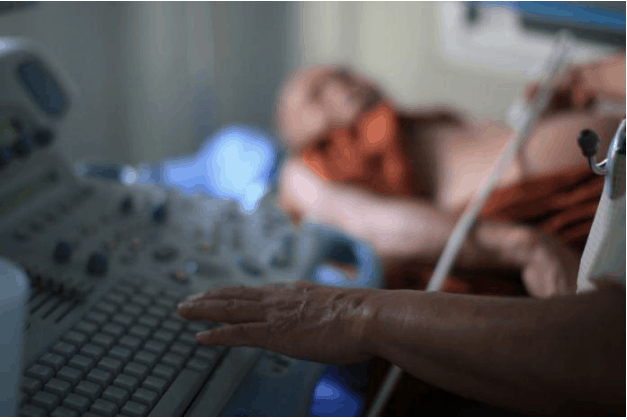
The most common type is the Transthoracic echocardiogram. This type is not painful in any way and is similar to an x-ray. It differs in that it uses sound waves to create the image and an x-ray uses radiation. Transthoracic ultrasound uses the same technology that doctors use to monitor babies in utero. A hand-held transducer probe connected to an ultrasound machine sends sound waves into the chest cavity. The returning waves create an image of the heart, and the doctor examines the images to make a diagnosis. Advances in ultrasound technology make it easier for doctors to see details in the images and make a more precise diagnosis.
During an echocardiogram, you will remove your shirt and get a hospital gown to wear. The sonographer will put three electrodes on your chest. They’re connected to an electrocardiograph monitor that will keep track of your heart’s activity. You’ll lie on your back, and the sonographer will place a transducer wand on your chest to get images of your heart. The process is completely painless and noninvasive. The only discomfort you might feel is from the cold wand. The whole procedure should take 30-40 minutes.
If you keep up with your doctor visits and follow their prescribed treatment for any issues related to your heart, you’ll live well into your golden years.
Senior Outlook Today is your go-to source for information, inspiration, and connection as you navigate the later years of life. Our team of experts and writers is dedicated to providing relevant and engaging content for seniors, covering topics such as health and wellness, finances, technology and travel.


